Foot & ankle pain
Overview
Our feet are a robust, complex and critical structure of our bodies. They bear our weight when we are moving and help ensure we can continue to move effectively and without pain. The foot contains 28 bones, 30 joints and more than a hundred ligaments, tendons and muscles that allow for a wide range of movement. Three of these bones – the tibia, fibula and talus – make up the ankle joint, and enable the up and down motion of the foot.
Foot pain is defined as discomfort felt in the toes, heels, arches, soles or other parts of the foot. It is an issue experienced by most people at some stage of their life, yet should not be considered normal. Although sometimes foot pain is only short lived, on occasion it continues for an extended period and when this occurs you should seek help from a podiatrist.
Foot pain is typically caused by a number of factors – lifestyle issues such as wearing unsuitable shoes, becoming overweight or pounding the feet on hard surfaces, medical conditions like diabetes, or an injury.
The ankle is the joint connecting the foot and leg, and is surrounded by a group of ligaments and tendons. It is a part of the body that absorbs a lot of stress and is prone to injury.
The most common foot and ankle conditions are diabetic neuropathy, bunions, ingrown toenails, metatarsalgia, morton’s neuroma, stress fractures, flat feet, plantar plate injury, sesamoiditis, posterior tibial tendonitis and arthritis.
You can find more information about the most common forms of foot and ankle pain below.
Diabetic neuropathy
Overview
Neuropathy is a term used to describe a complication of a number of different medical conditions. It involves damage to one or more of the nerve types responsible for sensation, power, movement and bodily functions performed by the gut, bladder and sweat glands. Most cases of neuropathy are found in people with diabetes.
Diabetic neuropathy is the occurrence of neuropathic symptoms in a person with diabetes. If you suffer from type 1 or type 2 diabetes, this condition is something you should be aware of. Diabetic neuropathy can affect your feet, and without proper care this condition can lead to injuries in the feet that may develop into infected sores and foot ulcers. This can affect a person’s quality of life significantly and in severe cases amputation of an affected limb may be necessary.
Symptoms
Up to 50% of people with diabetic neuropathy have no symptoms at all. Others may experience symptoms that don’t seem serious but develop slowly over months and years. It is very important for people with diabetes to undertake regular medical checks to look for early signs of this condition. Some of the signs of diabetic neuropathy may include one or more of the following:
- Numbness or reduced feeling in the feet.
- Tightness or burning, shooting, stabbing pains in the feet, hands or other body parts.
- A reduced or increased sensitivity to light, touch or temperature.
- Weakness and loss of balance and coordination.
- Extreme sweating on the torso, face or neck at night or when eating certain foods (spicy foods or cheese for example).
- Problems emptying the bladder properly or sensing when the bladder is full.
- Pain in and around the eyes, double vision and trouble moving an eye.
Causes
The high blood sugar levels common in people with diabetes can cause damage to blood vessels that supply oxygen and nutrients to the nerves in our feet. This can cause damage to the skin and the subsequent sensation, therefore making the feet more susceptible to damage. The following risk factors have also been shown to contribute to a diabetic person’s likelihood of developing diabetic neuropathy:
- Obesity.
- Smoking.
- Being older than 40 years of age.
- Extended periods of poor blood sugar level control.
- The chances of developing this condition increase the longer a person has diabetes.
- Hypertension.
- Heart disease.
Diagnosis
To establish if diabetic neuropathy may be present, a podiatrist would typically ask questions about your symptoms and health history. A physical examination would be conducted checking the sensation in your feet, the circulation, looking at the skin of the feet thoroughly and checking the tendon reflexes. It may be necessary for you to schedule a doctor’s appointment for more extensive testing based on the results.
Treatment
There is no cure for diabetic neuropathy, but controlling your diabetes properly and consistently is the best form of treatment. It is important to always keep your blood sugar levels within your target range to reduce symptoms.
It is also important to properly care for your feet. Diabetic neuropathy can cause the loss of feeling and sensation in the feet, increasing the risk of getting an unnoticed cut or scratch on your foot that can develop into a sore or ulcer, leading to the chance of serious infection.
Other treatments that may help to manage symptoms include:
- Regular exercise.
- Weight reduction.
- Enjoy a healthy and balanced diet.
- Quit smoking.
- Control your blood pressure.
- Limit or avoid alcohol consumption.
Prevention
Dan Everson Podiatry would recommend the following steps to help prevent diabetic neuropathy:
- Never walk barefoot.
- Check your feet daily for redness, swelling, cuts, bruises, sores, ingrown toenails, splinters and blisters using a mirror or ask a friend or family member to check for you.
- Schedule 12 monthly check ups with your podiatrist. Have them check for any corns or callouses on your feet. If you find a corn or callous prior to a checkup, do not use over the counter corn pads or razor blades.
- Wash your feet daily and ensure they are completely dry, paying particular attention to washing and drying the soles of your feet and in between the toes.
- Cut your toenails straight across with good quality nail clippers. File the nails afterwards.
- Moisturise your feet daily but do not apply lotion in between the toes.
- Wear seamless socks or stockings that are not too tight or restrictive.
- Ask your podiatrist about the best type of footwear and orthotic devices for your particular needs.
- Keep your feet away from direct heat from heaters, hot water bottles, scalding hot showers or baths and electric blankets.
- Exercise regularly but carefully to prevent injuring your feet.
Diabetic angiopathy
Diabetic Angiopathy is a serious disease of the blood vessels and is a common complication of chronic diabetes. It involves the decaying of the blood vessels and capillary tubes. There are two forms of Diabetic Angiopathy – micro-angiopathy which impacts the heart vessels and lower body limbs and macro-angiopathy which affects the eyes and kidneys. The triggers for diabetic angiopathy are the hormonal and metabolic disorders which stem from diabetes. Other risk factors include high blood pressure, smoking and regular alcohol consumption.
Symptoms of this condition include fatigue after exercise, and pain in the thighs and hips. This is followed by more acute pain in the feet when a person is lying down, which subsides during standing. At the condition’s most severe stage, the foot can become covered in ulcers and affected by gangrene. Diabetic patients should be checked regularly for diabetic angiopathy, and should immediately see their podiatrist if they suspect they have symptoms.
Bunions
A bunion is a bony lump or deformity at the joint of the base of the big toe. A bunion can occur when the joint at the base of the big toe becomes deformed and develops at a sideways angle, pushing the big toe inwards towards your other toes and sometimes displacing the long bones in the forefoot (the metatarsals). This condition is one of the most commonly seen by podiatrists.
Bunion sufferers will find it difficult to walk without pain. Thickening of the skin and tissues also occurs next to the base joint of the big toe which further contributes to the discomfort. A bunion will not go away of its own accord and will get worse over time, which in some cases can lead to permanent deformity and disability.
Symptoms
A person with a bunion may notice pain around the area and difficulty and increased pain when walking or running. The big toe will start to angle inwards towards your other toes – this can affect the second toe and cause it to become displaced also.
The area may become inflamed, swollen and painful to touch, with a lump of thickened and inflamed skin often appearing around the affected joint. Over time the foot may become too wide to fit into regular footwear. Arthritis of the big toe may also develop as a result.
Causes
Although it can be difficult to determine the exact cause of a bunion, it is thought certain hereditary foot types contribute to a large number of cases. These foot types have a faulty mechanical structure or characteristic making the person more likely to develop a bunion.
Other contributing factors include conditions affecting the joints such as osteoarthritis and rheumatoid arthritis.
Diagnosis
Dan Everson Podiatry can diagnose a bunion through a physical examination of the foot.
Treatment
There are several ways Dan Everson Podiatry would typically recommend, alone or in combination, to treat a bunion:
- Wearing appropriate and well-fitted footwear is essential to avoid further aggravating the condition. Roomy shoes will help to avoid rubbing of the bunion when you walk or run. Closed shoes, high heels and pointed shoes should be avoided.
- Following diagnosis, Dan Everson Podiatry may prescribe Kinetic Orthotics to insert into your footwear to help straighten the affected toes and treat other contributing factors. This is a range recommended by Dan Everson Podiatry, and is designed using patent-protected technology that optimises the way force is transferred as you move.
- Placing padding over the bunion will help to cushion it and prevent further rubbing and reduce impact to the area.
- Applying ice to the affected area during rest periods.
- Using pain-relief medication.
Prevention
If you think you may be at risk of developing bunions, Dan Everson Podiatry would recommend seeing your podiatrist for advice on what kind of footwear is appropriate for you.
Ingrown toenails
Overview
An ingrown toenail results when the toenail grows into the nail fold. It is a common condition experienced by most people at some stage and most often occurs in the big toenail, although any toe can become ingrown.
Symptoms
Ingrown toenails are usually quite painful, with the skin next to the nail becoming tender, swollen or overly firm. A usual sign is when the pain becomes worse when pressure is placed on the toe. If the toe becomes infected, people can experience more severe pain, pus leaking from the area and an overgrowth of skin around the toe.
Causes
The most common causes of ingrown toenails are:
- Poorly fitting shoes that are too tight or lose, placing extra pressure on the toe.
- Incorrect nail trimming – most often if the nail is trimmed too short, or if the edges are cut in a curved shape rather than straight across.
- Picking at the corners of the nails.
- A genetic disposition causing the nail to grow too thick, wide or curved for the skin around it.
- Trauma to the area resulting from stubbing your toe or other injuries.
- Feet that sweat a lot, leading to weak skin more easily pierced by the nail.
Diagnosis
A podiatrist can easily diagnose a case of ingrown toenails through a simple physical exam of the foot area.
Treatment
People with diabetes, nerve damage in the leg or foot, poor blood circulation or a nail infection who believe they have an ingrown toenail should consult with their podiatrist immediately. The condition can easily return without responsible foot care.
Below is a selection of the methods Dan Everson Podiatry would typically recommend, alone or in combination, for patients with ingrown toenails:
- Cutting back a wedge or a small section of nail called a spicule can provide instant pain relief.
- Consult with your GP for antibiotics may be required.
- Returning to see your podiatrist regularly to retrain the nail to grow normally.
- Thinning down the nail so it is less likely to pierce the skin.
- Nail surgery called ‘nail wedge resection’ may be necessary in chronic cases. This can be quoted and performed by Dan Everson Podiatry. It is a simple procedure with no stitching and minimal post-surgery pain, and has a success rate of 90%. It is covered by many private health insurers.
Prevention
Dan Everson Podiatry recommends the following activities to help prevent ingrown toenails.
- Trim your toenails straight, not curved.
- Don’t cut your toenails too short, as the pressure from your shoes can push a nail to grow into the skin.
- Ensure your shoes don’t pinch or put uncomfortable pressure on your toes. Dan Everson Podiatry can provide advice on the best style of shoes to look for.
- Soak feet in warm water to soften the nail before cutting.
- Use only clean, sharp nail trimmers.
- Avoid picking your toenails.
- People living with diabetes should have regular foot examinations and practice safe nail care.
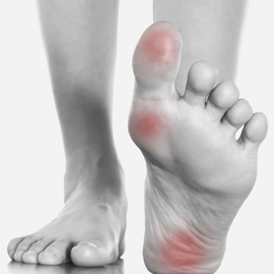
Metatarsalgia
Overview
Metatarsalgia is a term used to describe pain and inflammation in and around the long bones at the ball of the foot and can often include swelling around the joints in this part of the foot. These bones and joints absorb our weight when we walk and move. If damaged or weakened they can become painful and dysfunctional, causing discomfort and restricted movement.
It is also commonly referred to as a “stone bruise”, and it can be debilitating if left untreated. You may notice the pain appears gradually over several weeks.
Symptoms
People with metatarsalgia are likely to experience pain around the area of the forefoot, particularly at the ball of the feet and the toes. This has been described as feeling like you are walking on pebbles. Pain, general aching, burning or tingling sensations in the toes and tenderness of the forefoot area may increase when standing, walking and running. The general area at the ball of the foot will most likely feel tender when pressed.
Causes
Some of the more common causes of metatarsalgia include:
- Overuse and repetitive stress from activities such as running and jumping, commonly seen in most sports and made worse when performed on hard surfaces.
- Wearing high-heeled shoes and footwear that does not fit properly or support the foot appropriately.
- Being overweight. The metatarsals absorb the weight of our body when we are on our feet. The heavier we are, the more weight these bones are forced to support.
- Stiffness or limited movement in the ankle or Achilles tendon, causing the pressure of our bodyweight to be distributed unevenly across the foot.
- Having a very high arch in the foot can place excessive pressure on the ball of the foot.
- Morton’s neuroma – a condition that affects one of the nerves running between the bones in the forefoot causing pain, numbness and tingling sensations between two of the toes of the foot.
Diagnosis
An X-ray, bone scan or ultrasound can be used to show any problems with the bones, joints and soft tissue in the foot.
Dan Everson Podiatry can diagnose a case of metatarsalgia through history taking, a physical examination of the foot and a biomechanical assessment to study your range of movement. It may also be necessary to undertake a blood test to identify if diabetes, arthritis or gout is present. An X-ray, bone scan or ultrasound may also be advised by your podiatrist.
Treatment
Below is a selection of the methods Dan Everson Podiatry would typically recommend, alone or in combination, for patients suffering from this condition:
- Apply ice to the affected area for 15 – 20 minutes two or three times a day.
- Use pain-relief medication.
- Avoid running, jumping and other high impact exercises.
- Rest your feet and keep them elevated as much as possible.
- Wear shoes that are well fitted with a low heel. Kinetic Orthotics can be prescribed and inserted into your footwear to balance the foot and reduce stress on the metatarsals and forefoot. Dan Everson recommends its range of Kinetic Orthotics, which is designed using patent-protected technology to optimise the way force is transferred as you move.
- Footwear that doesn’t put pressure on the neuroma.
Prevention
Dan Everson Podiatry recommends the following steps to help prevent metatarsalgia:
- Ensuring you have appropriate footwear that distributes your weight evenly across the foot is very important.
- Massaging of the foot can be effective at the first sign of pain to relieve swelling pressure and increase blood flow to the area.
- Orthotics may help prevent this condition.
- Keeping your weight within a healthy range
- Walking and running on flat surfaces as much as possible.
Morton’s neuroma
Overview
Morton’s neuroma is a painful condition affecting the ball of the foot where the tissue tightens around some of the nerves leading to the toe, resulting in the nerve becoming swollen and irritated. It occurs most often in the area between the third and fourth toe, although it occasionally impacts the area between the second and third toe. Morton’s neuroma is more common in women.
Approximately 75% of people with Morton’s neuroma make a full recovery with appropriate care.
Symptoms
The main symptom experienced by people with Morton’s neuroma is a clicking feeling in the forefoot, followed by a sharp and burning pain or sensation of pins, numbness or needles extending to the end of their toes.
These symptoms typically become worse over time, and can be heightened through wearing narrow fitting shoes or extended periods of standing or walking. People with Morton’s neuroma can limp from the pain or feel forced to stop walking altogether.
Causes
Morton’s neuroma is most often brought about when the toes are squeezed together too often and for too long through activities such as wearing high heels or tight shoes.
Other contributing factors to Morton’s neuroma are:
- Being overweight.
- The ageing process that can weaken the pads that protect the feet.
- High impact exercises on hard surfaces that force the feet to absorb more impact from movement than they should.
- Abnormal foot movements used to ease the symptoms of bunions, hammertoes and flat feet.
- Having a stiff ankle or Achilles tendon.
Diagnosis
A podiatrist can diagnose a case of Morton’s neuroma through history taking and an investigation of your symptoms. Your feet will also be examined and a hands on examination will take place where your podiatrist will try to bring about your symptoms. You may also be encouraged to perform certain movements to help your podiatrist assess your foot alignment and function.
A blood test, foot X-ray, bone scan, ultrasound or MRI scans are sometimes required to firmly establish the cause of the pain.
Treatment
It is imperative to treat this condition immediately as the pain can spread to the rest of the foot and legs, eventually impacting a person’s ability to move freely.
Dan Everson Podiatry will recommend a treatment option based on the nature and severity of your condition. This may include:
- Rest and modifying your lifestyle to avoid unnecessary pressure on the feet while the area heals. This can involve using crutches and avoiding high impact exercises.
- Applying ice to the affected area for 15 minutes two or three times a day.
- Massaging the area.
- Anti-inflammatory drugs.
- Kinetic Orthotics are recommended by Dan Everson Podiatry to help ensure correct movement of the metatarsal bones to reduce compression on the nerves. Kinetic Orthotics are designed using patent-protected technology that optimises the way force is transferred as you move.
Prevention
Dan Everson Podiatry recommends the following activities to help prevent Morton’s neuroma.
- Warming up thoroughly before intense exercise.
- Stretching and strengthening the feet through exercises prescribed by your podiatrist.
- Ensure your weight sits within a healthy range to avoid stress on the feet.
- Avoid lacing the forefoot part of your shoes too tightly.
Metatarsal stress fracture
Overview
A Metatarsal stress fracture is a common injury impacting athletes and is caused by an incomplete crack in one of the metatarsal bones of the forefoot. The metatarsal bones are the five long bones in the front of the foot that connect to the toes.
Weight bearing activity places a load through the metatarsal bones. When the pulling forces placed on these bones through the attaching muscles becomes excessive, damage to the bones can gradually occur or can be caused by a sudden injury that can result in a metatarsal stress fracture. The second metatarsal (closest to the big toe) is most often impacted, as this is the bone that absorbs the most pressure during movement.
Metatarsal stress fractures are often caused by contact sports, and can be experienced by athletes, dancers and runners. With appropriate care management, most people recover fully from a fracture of this type within three to nine months.
Symptoms
A person with a metatarsal stress fracture will often experience a progressive pain in the front foot that gets worse when undergoing activities which bear weight on the foot such as walking and running. Swelling and tenderness around the area is often also experienced, with a sharp pain felt when pressure is applied to the metatarsal
.Causes
The most common causes of this condition are:
- Excessive weight bearing activities such as running, sprinting, jumping or dancing.
- A second toe being longer than the first.
- Poor quality footwear that doesn’t provide enough support.
- Landing on a hard surface from a height.
- The foot being stepped on or kicked, or having something dropped onto it.
- Not allocating enough rest time between activities or suddenly increasing the intensity of their exercise programs.
- When the foot rolls inwards excessively during walking or running, which results in the lower leg also turning inwards.
- When the metatarsal bones weaken due to conditions such as osteoporosis.
- When nerve sensation in the feet is weakened due to neurological issues such as diabetes.
- Tightness in the calf muscles.
Diagnosis
Dan Everson Podiatry can diagnose a metatarsal stress fracture through an examination of the area in conjunction with a bone scan that will provide a precise image of the fracture and can confirm the injury in very early stages. X-rays may be recommended for severe cases or at a later stage of the injury. MRI scanning is occasionally used to diagnose this injury.
Treatment
First and foremost, people with metatarsal stress fractures must ensure they rest the area as much as possible. Your podiatrist will guide you as to the degree of rest required, depending on the nature and severity of your fracture. Below is a selection of other treatments Dan Everson Podiatry would recommend for this condition:
- Stick to low impact activities such as swimming, bike riding or running in the water during the healing process.
- Crutches to avoid weight bearing on the affected foot.
- Anti-inflammatory medications.
- A cast or brace for severe cases where a fracture or displacement is likely.
- Protective footwear with more supportive soles to relief stress to the area.
- Surgery is rare but occasionally needed where a screw is inserted into the bone to help it heal.
When the rest period determined by your podiatrist has been completed and you have remained free of pain for a fortnight, you are able to consider a gradual return to a reduced movement exercise program. Keep in mind the foot will be particularly vulnerable for the first four weeks while it repairs itself.
Prevention
Dan Everson Podiatry recommends the following activities to help prevent metatarsal stress fractures.
- Custom orthotics can help prevent this condition through reducing the strain on the metatarsal bones. Dan Everson Podiatry recommends its range of Kinetic Orthotics, which is designed using patent-protected technology that optimises the way force is transferred as you move.
- Wear a supportive shoe at all times with appropriate arch support.
- Athletes should speak with their podiatrist about whether any modifications to their training program are needed.
Flat Feet
Overview
Flat feet is a condition in which the arch of the foot has not developed normally and is unusually low or flat. Several tendons of the foot and lower leg typically work together to form a foot’s arch. Flat feet can occur in one or both feet.
It is normal for babies and toddlers to have flat feet as the foot’s arch normally develops between the ages of three and five, yet some people never develop arches and others experience a fallen arch as they get older.
Symptoms
Some people with flat feet present with no symptoms and do not require treatment. Yet others may experience the following:
- The feet rolling in too much while standing or during movement.
- Aches and pain in the arches and heels of the foot, with pain heightening during activity.
- Challenges performing certain movements, such as standing on your toes.
- Back and leg pain.
- Swelling on the inside bottom of the feet.
Causes
Many people inherit flat feet, but occasionally the condition can result from:
- The connective tissue in the foot becoming stretched or irritated through overuse, inappropriate footwear, injury, ageing, rheumatoid arthritis or obesity.
- Conditions such as cerebral palsy, spina bifida or muscular dystrophy that affect the muscles and nerves.
Diagnosis
Dan Everson Podiatry will diagnose cases of flat feet through checking your health history and performing a biomechanical assessment during which your style of movement will be studied. The soles of your shoes may also be studied for insights into your wear patterns.
Treatment
If a case of flat feet is resulting in pain and restricted movement, Dan Everson Podiatry may suggest one or more of the following methods:
- Kinetic Orthotics may be prescribed to help treat flat feet through altering the neuromuscular function of the foot, and protecting it from high impact activities. This is a range designed by Dan Everson Podiatry, and is designed using patent-protected technology that optimises the way force is transferred as you move.
- Stretching exercises and various forms of physical activity.
- Footwear modifications to ensure all shoes have strong arch support.
- Pain relief medications.
- Rest and ice to reduce swelling and pain.
- For cases where foot pain is severe, surgery may be recommended on the rare occasion. Dan Everson Podiatry can provide you with a recommended course of action.
Prevention
Dan Everson Podiatry recommends the following activities to help prevent flat feet:
- Ask your podiatrist about stretches to prepare you for activities that will be taxing on your feet.
- Be aware of factors that can make flat feet worse, such as diabetes, high blood pressure and being overweight.
- Seek advice from a podiatrist on the most appropriate footwear or footwear modifications for your needs.
- Avoid high impact sports and excessive running and jumping on hard surfaces.
Plantar plate injury
Overview
Plantar plate injury is one of the most common causes for pain experienced in the ball of the foot. It refers to damage of the strong supporting ligament structure of the toe, located on the ball of the foot. The plantar plate is a thick structure that provides a large amount of stability to the toe, cushions the foot during weight bearing activities and helps bring the toe to the floor when standing. The plantar plate attaches to the base of the toe and the metatarsal, a long bone that connects to the toe.
This condition is most often experienced by mature-aged women whose feet tend to roll in. Most people make a full recovery within a few months.
Symptoms
Plantar plate injuries often result in ongoing pain and swelling in the ball of the foot that can extend towards the toes. This pain is likely to persist despite changes to footwear and lifestyle adjustments. Other common symptoms of this condition include:
- Splaying or clawing of the toes.
- Swelling and redness impacting the top of the foot.
- A sensation of ‘walking on the bones of the foot.
- A second or third toe that seems to be shifting position.
- A toe not touching the ground when a person is standing.
Causes
The most common causes of plantar plate injury are:
- Excessive loads being placed through the forefoot, impacting the plantar plates.
- Footwear that doesn’t fit properly.
- A short first metatarsal.
- A long second or third metatarsal.
- Imbalances in the foot and biomechanical issues.
- Untreated pigeon toe.
- Arthritis of the big toe.
- Bunions that push on the second toe.
Diagnosis
Dan Everson Podiatry will diagnose a plantar plate injury by checking your health history and performing a biomechanical and physical assessment during which your style of movement will be studied, and tests will be performed check the integrity of the plantar plate. An X-ray, MRI or ultrasound may be recommended.
Treatment
If a plantar plate injury is not treated adequately, there is a risk the condition can become chronic with a chance of deformity.
Dan Everson Podiatry would typically suggest one or more of the following treatments methods:
- Rest and ice to reduce swelling and pain.
- Anti-inflammatory medication.
- Kinetic Orthotics may be prescribed to relieve some pressure from the strained areas of the feet.
- Footwear modification to avoid high heels and tight shoes.
- Surgery is advised in some cases to repair the plantar plate.
Prevention
Dan Everson Podiatry recommends the following activities to help prevent a plantar plate injury:
- Custom orthotics can distribute pressure from the problem area. Dan Everson Podiatry recommends Kinetic Orthotics.
- Shoes which offload pressure from the ball of the foot. Your podiatrist can recommend some styles and suppliers to suit your feet.
Sesamoiditis
Overview
Sesamoiditis is a common condition impacting the forefoot that occurs when the sesamoid bones become irritated or fractured. The sesamoid bones are tiny bones found on the underside of the foot under the big toe joint within the tendons that run to the big toe.
Because of where the sesamoid bones are located, this condition also results in the tendons around the bones becoming irritated. They have a pulley-type function, increasing the leverage of the tendons controlling the toe. The sesamoid bones are often described to function like a kneecap for the big toe joint.
Sesamoiditis is most often experienced by young people, runners and dancers. With an appropriate care plan, most people fully recover from sesamoiditis within several months.
Symptoms
People with sesamoiditis often experience a dull pain in the ball of the foot, right underneath the big toe joint. This pain typically does not improve without treatment and can heighten to a sharp throbbing sensation. The sensations can come and go and usually become worse when wearing certain kinds of shoes or during particular movements.
Other common signs of sesamoiditis include:
- Swelling, bruising and pain in the ball of the feet and surrounding area.
- An inability to move the big toe freely.
- Limping or walking differently compared to usual in order to compensate for the discomfort experienced. This can in turn result in pain to people’s knees, hips, lower back and other parts of the foot.
Causes
The major causes of sesamoiditis are:
- A sudden increase in activity levels or change of exercise type that places more pressure on the balls of the feet.
- Bony feet with a lack of padding to protect the sesamoid bones.
- Feet with high arches can transfer pressure to the balls of the feet during certain movements.
- Flat feet.
- Wearing high heels.
- When the big toe is suddenly pulled upwards.
- Footwear with narrow toe boxes and toe springs.
Diagnosis
Dan Everson Podiatry is able to diagnose cases of sesamoiditis through history taking, a physical examination of the area and a Biomechanical Assessment to study your style of movement. An X-ray may also be recommended.
Treatment
Dan Everson Podiatry would typically suggest one or more of the following treatment methods:
- A period of rest.
- Anti-inflammatory medication.
- Applying ice to the area after exercise or movement that causes pain.
- Your podiatrist may advise you to wear a different style of shoe that can take pressure off the area and allow the toes room to spread.
- Avoiding wearing high heels while the area heals.
- Strapping or taping the big toe during the healing process.
- Your podiatrist may recommend you wear a cushioning pad under the area for extra support.
- A short removable leg fracture brace or cast may be advised for serious cases.
- Certain exercises can support the recovery process.
- Surgery may be considered in some cases. Dan Everson Podiatry can advise you on the best approach.
Prevention
Dan Everson Podiatry recommends the following activities to help prevent sesamoiditis:
- Kinetic Orthotics may help prevent this condition by cushioning the sesamoid bones if you have some of the risk factors. This is a range recommended by Dan Everson Podiatry, and is designed using patent-protected technology that optimises the way force is transferred as you move.
- The correct form of footwear that allow the feet enough room to move and do not cramp the toes.
- Replacing athletic shoes at least every six months.
Posterior tibial tendonitis
Overview
Posterior tibial tendonitis is a very common foot and ankle problem, experienced when the posterior tibial tendon becomes irritated or damaged. The posterior tibial tendon is one of the most important tendons of the leg, connecting the calf muscle to the bones on the inside of the foot, and helps to form the inside arch of the foot.
The main function of this tendon is to hold up the arch and support the foot during walking. Once this tendon becomes damaged, the arch will slowly collapse as a result over a period of time.
This condition is more likely to occur in women, people over the age of 40 and those who are obese, and have diabetes or hypertension.
Symptoms
The tendon of people with posterior tibial tendonitis typically provides less stability and support for the arch of the foot. This can result in:
- Pain experienced on the inside of the foot and ankle.
- People feeling unsteady on their feet, experiencing muscle weakness or difficulty rising onto their toes on the side that is affected.
- Pain which gets worse with movement or long periods of standing.
- Pain experienced on the outside of the ankle. This occurs as a result of the heel bone repositioning itself and placing pressure on the outside ankle bone.
More serious cases can result in a flattening of the inside arch of the foot resulting in flatfoot, and toes ‘splaying’ outwards.
Causes
The most common causes of posterior tibial tendonitis are:
- Continued wearing of poorly fitting or unsupportive shoes.
- Trauma or injury to the ankle joint.
- Poor foot posture, with too much pressure placed on the ankle joint.
- Overuse of the tendon by athletes or physically active people.
Diagnosis
A podiatrist can diagnose a case of posterior tibial tendonitis through a physical exam of the foot and ankle area and an investigation of your symptoms. You may be asked to walk to help your podiatrist assess your range of motion and flexibility.
Your podiatrist may also recommend an X-ray, MRI, CT scan or ultrasound as part of the diagnostic process.
Treatment
Dan Everson Podiatry will recommend a treatment option based on the nature and severity of your injury. These may include:
- Rest and modifying your lifestyle to reduce or stop painful activities. Changing to low impact exercise is usually recommended.
- Ice therapy to the affected area three or four times a day to reduce swelling.
- Strength and conditioning work.
- Stabilisation (strapping, splitting, casting) may be recommended to allow the area to rest and recover.
- Surgery may be needed in some cases, which requires referral from a doctor to an orthopaedic surgeon.
- Anti-inflammatory medication.
- Orthotics may be prescribed to help treat this condition and prevent a recurrence. Dan Everson recommends its range of Kinetic Orthotics, which is designed using patent-protected technology that optimises the way force is transferred as you move.
Prevention
Dan Everson Podiatry recommends the following activities to help prevent posterior tibial tendonitis.
- Wear supportive shoes and customised orthotics to reduce stress on the tendon.
- Perform stretches daily that strengthen the calf and posterior tibial tendon.
- Balance work on a balance board or Bosu ball can alleviate stress to the tendon.
Arthritis of the foot and ankle
Overview
Arthritis is the inflammation of one or more of the joints and can impact the small joints and surrounding tissue of the foot and ankle, making it challenging to move without pain. The joints of the feet help enable a wide range of movement, and are often surrounded by a soft connective tissue called cartilage that helps bones glide smoothly over each other when a person moves.
Arthritis cannot be cured, however there are many care options available to treat the symptoms and slow its progress to help reduce pain.
There are three types of arthritis most likely to impact the foot and ankle – these are osteoarthritis, rheumatoid arthritis and posttraumatic arthritis.
Post-traumatic arthritis
Overview
Post-traumatic arthritis typically develops after an injury to the foot or ankle – most commonly dislocations and fractures. This form of arthritis also sees the cartilage between joints erode, which can take place many years following an injury.
An injured joint is seven times more likely to develop into arthritis compared with an uninjured joint, even if the injury is professionally treated.
Osteoarthritis
Overview
Osteoarthritis is the most common type of arthritis. It is a degenerative condition often experienced by middle-aged people. The cartilage in the joint disintegrates over time, becoming rough and minimising the protective space between the bones. As a result, the bones may rub together which causes the joint to become painful and inflamed.
In the foot, osteoarthritis most frequently impacts the big toe, although it occasionally occurs in the midfoot and ankle.
Symptoms
The condition develops gradually and results in pain and stiffness to the area that gets worse over time. People with osteoarthritis will usually find it difficult to walk, bear weight or bend the joint without pain. Bone spurs can also develop as a result of osteoarthritis.
Causes
The below are common causes of osteoarthritis:
- Obesity and a family history of the condition.
- An injury can lead to osteoarthritis in 15 percent of cases – such as kicking or jamming the big toe, dropping something on the midfoot, or a fracture of the ankle. This is called post-traumatic arthritis.
- Atypical foot mechanics – flat feet or high arches which cause strain on the joints.
Diagnosis
Dan Everson Podiatry can diagnose osteoarthritis through history taking, a physical examination of the foot and ankle and a biomechanical assessment to study your range of movement, look for swelling in the joint and pain experienced through movement. An X-ray, MRI or CT scan may also be recommended to evaluate the stage of the illness.
Treatment
Dan Everson Podiatry would typically suggest one or more of the following treatment methods:
- Anti-inflammatory medication.
- Kinetic Orthotics may be prescribed to help the foot move more freely or for cushioning support to provide pain reduction. This is a range designed by Dan Everson Podiatry, and is designed using patent-protected technology that optimises the way force is transferred as you move.
- Weight loss may be recommended for overweight patients.
- Shoe modification.
- A foot brace to protect the foot from movement and support the joint can reduce pain during walking and help prevent alignment problems.
- A cast to restrict movement while the inflammation reduces.
- Your podiatrist can recommend certain exercises to strengthen and stabilise the area, and minimise risk of injury.
- Surgery may be recommended in some cases with osteoarthritis has progressed to an advanced stage or when other forms of treatment have not improved the condition.
Prevention
Dan Everson Podiatry recommends the following activities to help prevent developing osteoarthritis in the foot and ankle.
- Control your weight to lessen strain on the joints.
- Avoid foot and ankle injuries by wearing appropriate shoes, avoid hard surfaces when exercising and land with the knees bent to improve shock absorption.
- Treat any joint injuries as quickly as possible.
Rheumatoid arthritis
Overview
Rheumatoid Arthritis is a chronic autoimmune disease that often impacts multiple joints of the foot and ankle. 90 per cent of rheumatoid arthritis patients will eventually experience symptoms in the foot and ankle.
This condition occurs when the immune system attacks its own tissues. The immune cells of people with this condition attack the soft tissue between the joint capsule and joint cavity of the synovial joints, which causes the area to become swollen. Over time, the synovium damages the bone, cartilage, ligaments and tendons and can cause joint deformity and disability. It mainly affects the lesser joints such as the metatarsophalangeal joints of the feet.
This form of arthritis affects 1% of the population, with women more than twice as likely to develop the condition than men.
Symptoms
Symptoms are most often experienced in the toes and forefeet first, then in the back of the feet, and then the ankles.
The symptoms most often associated with rheumatoid arthritis are:
- Deformities of the feet.
- Pain, swelling and stiffness of several joints in both feet. This typically manifests in pain in the joint, sole or ball of the foot.
- Development of corns or bunions on the feet.
- Toes that curl and stiffen.
- Bones that shift, causing the foot’s arch to collapse, resulting in flat feet.
- Fever, fatigue and appetite loss of appetite.
- Softening of the bone resulting in stress fractures and bone collapse.
- Difficulties walking up inclines and stairs.
Causes
The causes of rheumatoid arthritis are presently unknown.
Diagnosis
Dan Everson Podiatry can assess for rheumatoid arthritis through history taking, a physical examination of the foot and ankle and a biomechanical assessment to study your range of movement, look for swelling in the joint and pain experienced through movement. An X-ray, MRI or CT scan may also be recommended to evaluate the stage of the disease. A blood test through the doctor and consultation with a rheumatologist may also be required to diagnosis and manage in condition, in conjunction with podiatric care.
Treatment
Dan Everson Podiatry would typically suggest one or more of the following treatment methods:
- Anti-inflammatory, pain relief and prescription medication which can help slow the spread of the condition.
- Rest and applying ice to the area.
- Kinetic Orthotics may be prescribed to help the foot move more freely or provide pain reduction. This is a range designed by Dan Everson Podiatry, and is designed using patent-protected technology that optimises the way force is transferred as you move.
- Shoe modification – deep toe boxes or soft arch supports with a firm heel can be helpful.
- Canes and crutches may be advised for severe cases.
- A cast to restrict movement while the inflammation reduces.
- Your podiatrist can recommend certain exercises to improve your range of movement.
- Surgery may be recommended in some cases. The most common procedure performed for rheumatoid arthritis is the fusion of the affected joints.
Prevention
There is no known way to prevent rheumatoid arthritis, as the causes are presently unknown. There are several ways to reduce your risk of major joint damage after a diagnosis of this condition:
- Consult with your doctor as soon as you have any symptoms in order to be properly diagnosed, so you can be managed by your podiatrist appropriately.
- Speak with your podiatrist about a program of gentle stretches and strength movements to reduce pain and strengthen the bones.
- Rest from exercise during the periods you are experiencing the most pain.
- Avoid smoking.